Top-level research with nanolight
Extremely thin metal tips and optical fibres analyse the finest structures
A ›Turningtable‹ for nanomaterials: Teams of researchers from the fields of chemistry and physics are developing ultra-sharp metal tips and optical fibres to scan and analyse the finest surface structures. When combined with intense laser light, the metal tips become super-focusing nanolights that are able to detect various features of surface structures, such as their spatial dimensions, chemical composition and optical properties.
By Ute Schönfelder
The research teams directed by Prof. Dr Volker Deckert and Prof. Dr Thomas Pertsch are investigating light-matter interactions using a high-resolution optical method that incorporates nonlinear optical effects within the established principle of scanning probe microscopy. »To put it simply, a scanning probe microscope works like a Turntable,« explains Thomas Pertsch. A tiny metal tip scans the surface of the material sample and transmits information about its topography to a detector via a moving arm. »This method is so sensitive that metal surfaces can be resolved down to individual atoms in the metal lattice,« says Pertsch.
But that’s not sensitive enough for the physicists… As part of a collaborative project, the researchers are combining scanning probe microscopy with optical methods to obtain information on the optical and chemical properties of samples in addition to their surface structure. Volker Deckert and Thomas Pertsch have each been pursuing different approaches, which will be combined into a common measurement method at the ›NOA‹ collaborative research centre.
Deckert and his colleagues from the Institute of Physical Chemistry are using tiny silicon needles to scan the samples. The needles themselves are around 50 micrometres long, which is less than the thickness of a sheet of paper. The tip of the needle that interacts with the sample is over 1,000 times smaller, measuring just five to ten nanometres in diameter. But even that’s not small – and therefore sensitive – enough… »The silicon needles are also coated with silver vapour, which is deposited on the silicon surface as tiny droplets,« explains Deckert. The individual silver droplets increase the effective needle point only insignificantly by around 10 nm, which roughly equates to the thickness of a cell membrane.
By preparing the needles in this way, the tiny probes are still able to scan details of the sample, revealing the smallest intricacies of its structures and defects. In addition, once irradiated with laser light and coupled to a Raman spectrometer, the light scattered by the needles can also be used to detect the surface structure and its properties.
The main objective of the researchers, however, is actually something else: »The concentration of high-intensity light particles in the very small volume of the metal needles causes nonlinear effects between the light and material,« says Deckert. The needle tips become a source of light, so to speak, locally concentrating the light particles so strongly that the sensitivity of the Raman spectroscopy process is significantly enhanced. Similar to the team directed by Jürgen Popp and Jer-Shing Huang, Deckert is using linear and nonlinear light-matter interactions to coherently bundle the Raman scattering excited by intense short-pulse lasers (›coherent anti-Stokes Raman scattering‹), enhancing the Raman signals as a result.
In a recent study published in cooperation with their US colleagues, the researchers from the University of Jena demonstrated the highly practical potential applications of these methods. They presented their results in ›Proceedings of the National Academy of Sciences‹, showing that tip-enhanced Raman spectroscopy can be used to detect virus particles in clinical samples and even identify the respective virus type – all in the time of a few minutes. While the study focused on influenza and coxsackieviruses, the method would theoretically also be suitable for detecting other pathogens, such as SARS-CoV-2, emphasizes Volker Deckert.
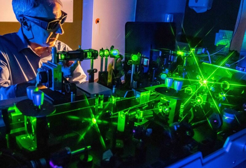
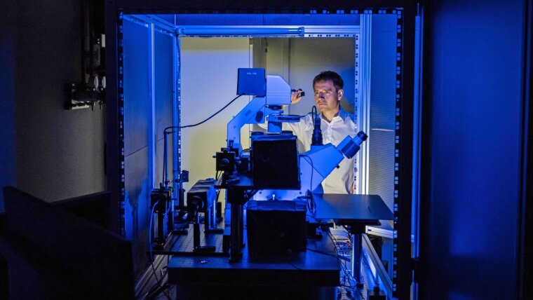
Prof. Dr Volker Deckert sets up a laser to examine nanoscale surfaces and their nonlinear optical properties.
Image: Jens Meyer (University of Jena)When it comes to producing light-focusing needles for nonlinear optical investigations, Thomas Pertsch and his team at the Institute of Applied Physics are taking a different approach: »We’re using optical fibres coated with a thin layer of gold at the tip,« explains Pertsch. The fibre itself is a ›kitten’s whisker‹, measuring 125 micrometres in diameter – and therefore slightly thicker than a human hair. Its tip is approximately 50 micrometres long and tapers to a few nanometres. When laser light passes through the fibre at the appropriate wavelength, the light particles engage in a resonant interaction with the gold coating’s electrons at the gold-plated tip. This creates a ›surface plasmon‹ – the collective excitation of electrons along a metal surface – which interacts with the light particles. »The light focused at the tip of the needle is practically trapped there due to the interaction with the electrons – it can’t escape,« says Pertsch. The sample can be scanned with the shining needle as it interacts with the light. At the same time, the fibre returns the light reflected by the sample and transmits it to a detector.
These methods are providing the researchers with extensive and rich information about the samples examined. »The data allows us to draw conclusions about the optical properties produced by each sample structure,« says Deckert, who adds that their eventual target is to produce nanostructures in a targeted manner so that the materials have the exact optical properties required for a specific application.
The aim of the current research project at the ›NOA‹ collaborative research centre is to carry out basic research to establish the investigation methodology on well characterized reference materials. »Another key ingredient of our success is our close cooperation with research partners who are creating theoretical models of light-matter interactions,« emphasizes Thomas Pertsch. This is essential both for the development of nanoscale tips and the evaluation of nonlinear optical effects.

Here we can see a silver-plated silicon tip used to scan surfaces and materials. The metal tip significantly increases the sensitivity of Raman spectroscopy.
Image: Jens Meyer (University of Jena)Original-Publication:
Laser spectroscopic technique for direct identification of a single virus I: FASTER CARS, Proc Natl Acad Sci U S A. 2020, DOI: 10.1073/pnas.2013169117External link